Vascular Surgeon
Expert in the treatment of varicose veins
Questions and answers about varicose and spider veins and their treatment

What are spider veins?
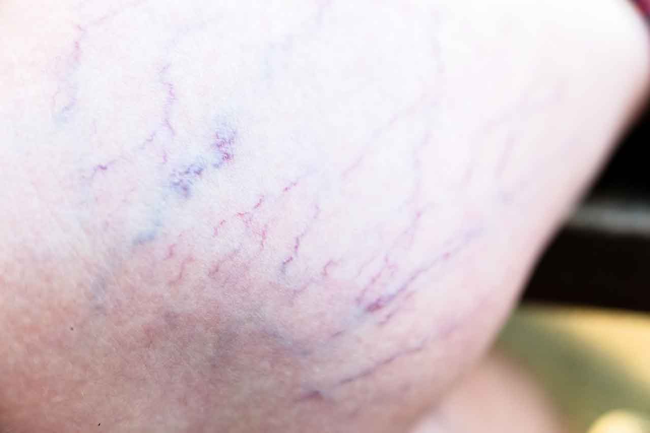
Spider veins is the common name for what are known in medical terms as "telangiectasias". This is the dilation of tiny vessels (veins or capillaries) located immediately below the epidermis (the most superficial layer of the skin).
Telengiectasias can occur anywhere on the body, most commonly on the face and legs. Vascular specialists generally focus on telengiectasias in the legs, while facial telengiectasias are more often managed by dermatologists.
How do you recognize them?
They form fine networks or circumscribed patches of varying size, forming a hair follicle in the form of a star, spider's legs or sometimes patches. They can be of different colors, from red to dark purple, and these different colors can co-exist on the same leg.
They can also vary in size, from hair-thin to larger varicose dilatations. One particular form is represented by what is known as "matting", which takes the form of a nebulous red spot, without the small vessels that make it up being identifiable.
There is in fact an almost infinite variety of forms and combinations of telengiectasias, but what they all have in common is that they are very superficial and disappear when pressed (disappearance with vitropressure).
How do they appear?
The pathophysiology of telengiectasias in the legs (their cause and risk factors) is still rather mysterious today.
They are certainly correlated with venous insufficiency, and therefore with its risk factors (heredity, age, chronic foot position, exposure to heat, pregnancy, female hormones), but what triggers their appearance is not clearly established. They represent the first stage of venous insufficiency, but they are very common (at least one in two people over the age of 50 are affected) and the majority of patients affected by them will never progress to the more advanced stages, in particular the "true" varicose vein stage.
Telengiectasias appear to be more frequent in women, suggesting a hormonal factor (estrogen-progesterone) in the permanent dilation of these small vessels. They are also more frequent with age, as aging certainly favors their appearance.
Trauma (shock, injection, operation) causing bruising on fragile skin could also be the cause of telengiectasias. It seems that certain skin types are more prone to the emergence of telengiectasias.
Is there any way to prevent telengiectasias?
It's very difficult to answer this question, because as we've just mentioned, we don't know exactly why they develop.
Preventing or limiting the appearance of telengiectasias is, in spite of everything, based on the rules for preventing venous insufficiency (physical activity, wearing venous compression, early treatment of pathological veins, combating the risk factors of venous insufficiency), since they represent the first stage of venous "pathology".
Preventing trauma can also play a role when telengiectasias are already present, or if there's a family history of them, by avoiding sports involving contact with the legs, and certain procedures that may involve skin trauma (pressure massage, palpate-roll, or any other procedure leading to bruising).
The question of the risk of taking hormonal treatments can be raised to a certain extent, by assessing the benefit-risk balance, as telengiectasias usually only cause an aesthetic problem.
While it's true that it's very difficult to prevent telengiectasias before they appear, it is possible to limit their development once they are present, particularly through early treatment.
Are any special tests needed for telengiectasias?
It is advisable to consult a vascular specialist, who, after examining the leg, will carry out an echo-Doppler scan to assess the venous circulation of the limb.
The presence of telengiectasias can sometimes reveal a disturbance of venous circulation in the larger vessels of the leg (saphenous veins and/or their branches), although in most cases this examination will be normal.
More specific examinations, such as transillumination, show the relationship between telengiectasias and the network of small veins.
Similarly, there are now devices that can visualize superficial vein networks directly on the skin. Vein detectors" use near-infrared light reflection to create a map of the veins, with the imagery recorded by the device then projected onto the patient's skin. While this technology is spectacular, it cannot distinguish "diseased" veins (those with reflux) from healthy veins (those that drain blood normally), unlike echo-Doppler examination.
These techniques for visualizing the venous network can be useful for guiding the treatment procedure.
Are telengiectasias risky? Can they cause complications?
Telengiectasias are not really a medical risk, as in the vast majority of cases they do not cause complications. However, telengiectasias can rupture, with the risk of hemorrhaging. This can be spectacular, but is always easy to stop by elevating the leg and compressing the bleeding point.
This complication is only seen in advanced cases of venous insufficiency, usually with significant involvement of the main superficial veins, and sometimes with skin lesions characteristic of advanced stages (hypodermatitis, varicose ulcers).
The occurrence of hemorrhage from ruptured telengiectasias is a clear indication for therapeutic treatment, to stop the hemorrhage or prevent recurrence, in contrast to treatments for ordinary telengiectasias, which are purely cosmetic.
How can telengiectasias be treated?
Sclerotherapy has been the reference treatment for over 70 years. The principle is to inject an irritant directly into the varicose vein, using a very fine needle. This causes inflammation of the vein wall, leading to its "destruction" and the ultimate stage of inflammation, that is sclerosis.
Polidocanol, sodium tetradecyl sulfate and chromated glycerin are the main liquid substances used, depending on country and habits. The first two can also be used in foam form (by mixing with air), although opinions differ as to the greater effectiveness of the foam form.
Depending on the extent of telengiectasias, several sessions are generally required, with intervals of 2 to 4 weeks. In most cases, the result is not definitive, and maintenance sessions are necessary to prevent the extension of new telengiectasias over time. This is an aesthetic treatment, except in cases of hemorrhage caused by ruptured telengiectasias (see above).
The other possible treatment, which appeared some thirty years ago, is cutaneous laser treatment. The principle is to apply a laser beam to the skin, targeting the telengiectasias, so that the laser light is absorbed by the telengiectasias (essentially by the hemoglobin pigments present in the vessel), destroying them through a thermal effect. Different lasers with different wavelengths are available, depending on the location and appearance of the telengiectasias.
Most vascular specialists use sclerotherapy in the first instance, as it is less costly and easier to perform. Laser treatment is generally reserved for cases resistant to sclerotherapy, but can be used for all types of telengiectasias in the hands of practitioners who have mastered the art.
There is no scientific consensus on the use of either technique. As with any treatment, there is a risk of side-effects with these techniques (pigmentation disorders, skin lesions, thrombosis), although serious complications are extremely rare.
Aesthetic results can vary, however, and are never certain and definitive.
In summary
Spider veins (or telangiectasias of the lower limbs in more medical terms) are a very common and benign condition, with rare exceptions.
Even if they only cause cosmetic discomfort, they represent the first stage of venous insufficiency.It is therefore necessary to consult a vascular specialist when they are present, in order to rule out the existence of any anomaly in venous drainage, the treatment of which would help prevent progression to a more advanced stage and facilitate treatment of the telengiectasias themselves.
When the functioning of the main venous system does not present any anomaly, which is the most frequent case, the treatment will be purely cosmetic.The treatment of telengiectasias is most often based on sclerotherapy, which requires the expertise of a vascular specialist, often requiring several sessions to obtain a satisfactory result, and the need for monitoring, as this treatment is not definitive.
For certain practitioners or in certain indications, cutaneous laser treatment is preferred, with the same principles of repeated treatment and medium- and long-term monitoring.


